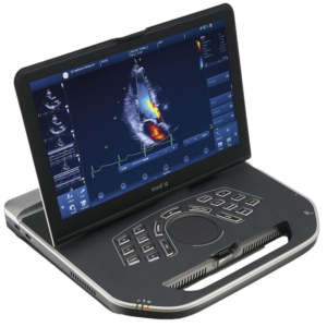
A Heart Sonar, also known as an echocardiogram, is a non-invasive imaging test that uses ultrasound waves to create detailed images of the heart’s structure, chambers, valves, and blood flow patterns. This diagnostic procedure provides valuable information about cardiac function and helps diagnose various heart conditions. Here’s a detailed explanation of what a Heart Sonar (Echocardiogram) is and why you might need one:
What Is a Heart Sonar (Echocardiogram)?
Ultrasound Imaging:
A Heart Sonar (echocardiogram) uses high-frequency sound waves (ultrasound) to produce real-time images of the heart.
A handheld device called a transducer is placed on the chest and emits ultrasound waves that bounce off structures in the heart, creating detailed images displayed on a monitor.
Types of Echocardiograms:
Transthoracic Echocardiogram (TTE): The transducer is placed on the chest wall to obtain images of the heart from the outside.
Transesophageal Echocardiogram (TEE) (Available only in MediClinic per special referral from Physician or Cardiologist): A specialized probe is inserted into the esophagus to obtain closer and more detailed images of the heart structures.
Why Might You Need a Heart Sonar (Echocardiogram)?
Diagnosis of Heart Conditions:
A Heart Sonar (echocardiogram) is used to diagnose various heart conditions, including:
Valvular Heart Disease: Evaluates the structure and function of heart valves (e.g., mitral valve prolapse, aortic stenosis).
Cardiomyopathies: Assesses the size, shape, and function of the heart muscle (e.g., hypertrophic cardiomyopathy, dilated cardiomyopathy).
Congenital Heart Defects: Detects abnormalities present at birth (e.g., atrial septal defect, ventricular septal defect).
Pericardial Disease: Evaluates the pericardium (the sac surrounding the heart) for fluid accumulation or inflammation.
Assessment of Cardiac Function:
Echocardiography measures parameters such as ejection fraction (EF) to assess the heart’s pumping ability.
It evaluates cardiac chambers’ size, wall thickness, and motion abnormalities.
Monitoring Cardiac Health:
Regular echocardiograms are performed to monitor disease progression, treatment response, or recovery after cardiac procedures (e.g., heart surgery, valve replacement).
Evaluation of Symptoms:
If you experience symptoms such as chest pain, shortness of breath, palpitations, or unexplained fatigue, an echocardiogram helps determine the underlying cause.
Importance of a Heart Sonar (Echocardiogram):
- Non-Invasive Imaging: Echocardiography is safe, painless, and does not involve radiation exposure, making it suitable for repeated evaluations.
- Accurate Diagnosis: It provides detailed information about cardiac anatomy and function, aiding in the accurate diagnosis and management of heart conditions.
- Guiding Treatment Decisions: Echocardiographic findings guide treatment strategies, including medication management, surgical interventions, or implantation of cardiac devices (e.g., pacemaker, implantable cardioverter-defibrillator).
In summary, a Heart Sonar (echocardiogram) is a valuable diagnostic tool used to assess heart structure, function, and blood flow patterns. This non-invasive imaging test plays a critical role in diagnosing heart conditions, guiding treatment decisions, and monitoring cardiac health to optimize patient care and outcomes. If you have symptoms suggestive of a heart problem or require routine cardiac evaluation, your healthcare provider may recommend a Heart Sonar (echocardiogram) to assess your heart’s health comprehensively.
How long does a Heart Sonar test take, and is it safe?
A Heart Sonar typically takes about 30-60 minutes. It’s a safe and painless procedure that doesn’t involve radiation. The images produced are valuable for diagnosing various heart conditions.